Spotting a mole on your dog that seemingly appeared out of nowhere can be really scary.
And believe me, when I found one on my six-year-old female Rottweiler, I was concerned.
It just happened while casually stroking her, like I would every day, and then I felt a slight bump beneath her fur.
In itself, a mole is not a big problem as lumps, bumps, and cysts commonly happen in older dogs so we decided to wait and observe it for any changes.
If you are in the same position make sure to track the mole’s progress by taking pictures at least once a week.
Unfortunately, the mole did change and quickly doubled its size after a few weeks so I quickly started researching what this could mean.
When to Worry About a Black Mole on Your Dog
Finding a black mole on your dog can be concerning, but not all moles are cause for alarm. Dogs, like humans, can develop skin growths for a variety of reasons, many of which are benign.
However, there are some warning signs that may indicate the mole needs further attention from a veterinarian.
1. Changes in Size, Shape, or Color
One of the biggest red flags is if the mole starts to grow rapidly, changes shape, or darkens significantly over a short period.
These changes could indicate that the mole is not a simple skin growth but potentially a sign of a more serious condition, such as a tumor.
Our dog’s mole grew from a small pigmented area to 1cm in diameter within three months.
2. Irregular Edges or Asymmetry
Healthy moles are usually symmetrical and have smooth, even edges.
If the mole appears irregular in shape or has jagged edges, it’s worth getting it checked out.
Asymmetry, where one half of the mole doesn’t match the other, can also be a warning sign.
3. Bleeding, Itching, or Discharge
A mole that starts to bleed, ooze, or cause your dog discomfort (e.g., excessive itching or licking) is not normal.
These symptoms could suggest irritation, infection, or even malignancy and should be evaluated by a vet as soon as possible.
4. Location and Interference
Sometimes, the mole itself may not be dangerous but can be problematic based on its location.
For example, a mole that your dog frequently scratches, bites, or rubs against surfaces might become irritated or infected.
Even benign moles in these situations might need to be removed for your dog’s comfort.
5. Your Dog’s Age and Breed
Certain breeds, such as Boxers, Golden Retrievers, and Labrador Retrievers, are predisposed to developing skin tumors.
Additionally, older dogs are more likely to develop malignant growths. Knowing your dog’s risk factors can help you determine whether a mole is worth closer examination.
Some common benign skin growths include warts, skin tags, cysts, and benign tumors. However, most of these are actually not black in color.
Just like in humans, a black mole could indicate a highly aggressive skin cancer called melanoma and this was our biggest concern.
When researching on the topic of melanomas in dogs, I found that, unlike humans, dogs can have benign melanomas and they are diagnosed much more frequently.
I discovered that in dogs, melanomas have a 70%1 to 80%2 chance of being benign.
And whether they are benign or not largely depends on the position.
The most aggressive and dangerous type of melanoma in dogs is oral melanoma (80%3).
This form of melanoma typically occurs in the mouth, gums, or around the lips, and it has a high potential for malignancy.
Oral melanomas are considered to be the most lethal form of canine melanoma, with a reported median survival time of just 65 days in dogs left untreated.
Treatment of Canine Oral Melanomas: A Critical Review of the Literature
Other dangerous types include digit (toe) melanoma and mucosal melanoma.
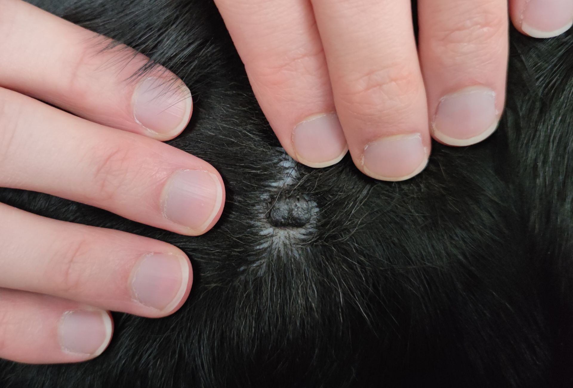
Amalia’s mole is located on the right side behind her shoulder on haired skin and cutaneous (skin) melanoma have the highest likelihood of being benign.
These tumors are usually non-invasive and do not spread to other parts of the body.
Here are a few images that I took over the course of a few weeks:

Due to the rapid growth, the black color, and its being raised, we decided to take her to the vet.
Dog Mole Diagnosis
Diagnosing a skin growth in a dog typically involves several steps performed by a veterinarian to determine the nature of the growth and whether it poses any health risks.
The veterinarian will start by visually examining the growth, and assessing its size, shape, color, texture, and location. They will also check for any signs of irritation, ulceration, or discharge.
During this examination, they may palpate the growth to determine its consistency (soft, firm, or hard) and whether it is attached to underlying tissues.
Our vet determined that it did look suspicious and wanted to take further steps.
We had already decided that we definitely wanted to test the skin growth to have a definitive result.
There are two main ways to take tissue sample from a skin growth – the fine needle aspiration and a biopsy.
A fine-needle aspiration is a minimally invasive procedure where the vet uses a small needle to collect a sample of cells from the growth.
These cells are then examined under a microscope to identify their nature (e.g., benign, malignant, or inflammatory).
The advantage of this technique is that your dog won’t require an aesthetic however it sometimes happens that not enough cells have been collected for a conclusive result.
If the fine-needle aspiration results are inconclusive or suggest malignancy, the vet may recommend a biopsy.
This involves removing a larger sample of the tissue, or sometimes the entire growth, for histopathological examination by a veterinary pathologist.
A biopsy provides the most definitive diagnosis.
We decided on completely removing the whole mole via surgery for one simple reason.
Even if a biopsy comes back with a positive result, a benign mole can always turn malignant at any point in the future.
When it’s fully removed it has a very small chance of reoccurring.
Dog Mole Removal Surgery
Removing a mole on a dog surgically is usually a very short operation under general anesthesia.
Our dog had to fast for 12 hours and was not allowed to drink water for 2 hours pre-surgery.
On Monday, we dropped her off at the vet for her procedure. She received an initial injection in her bottom to help calm her, and then she was carried to the operating room where she was given intravenous (IV) sedation.
Because the procedure was relatively short, the vet opted not to use inhalation anesthesia (which is otherwise recommended).
The surgery itself, which included the mole removal and a thorough teeth cleaning, took about 30 minutes. We were able to pick her up roughly an hour later, once she was fully awake.
For the rest of the day, she was understandably drowsy and slept most of the time. In the evening, we offered her a small portion of food, which she eagerly devoured.

She was absolutely starving by then, and waiting all day for her first meal was probably the most challenging part of the experience for her.
Thankfully, she had no side effects from the anesthesia. The vet provided us with some pain medication for the following days and she had already received a dose during the surgery itself.
That first night was tough. She was restless, whining and shifting around, and it took some time before she finally settled down. She only relaxed once we invited her onto our bed.
This is what the incision looked like one day post-surgery:

By Wednesday, we had her post-operation check-up. The vet confirmed that her wound was healing beautifully, which was a huge relief.
Caring for Your Dog After Mole Removal Surgery
One thing we learned the hard way: if your dog ever needs skin surgery, invest in an operative body suit beforehand.
We underestimated how much she would try to scratch at her stitches, and it required constant supervision to stop her.
We ordered one right away, but due to holiday delays, it didn’t arrive before Christmas. Ironically, it’s likely to show up just in time for her stitches to be removed.
The other thing that really helped was securing the one paw that could reach the incision site with a long and thick sock.
That way she wouldn’t be able to do much harm if she decided to scratch the site and we still monitored her closely.
Originally the site was covered by a bandage which was supposed to be removed on the post operation check up however she managed to remove it within the same day.
Luckily, we had some other bandages prepared that worked just as well.
After that, the incision should be left uncovered and dry. We were not told to put anything on it and to just leave it be.
She didn’t develop any redness or swelling around the area, however on day six a small area turned pink but it cleared up during the night.

After 11 days, the stitches were removed and the incision continued to heal well after that.
Our Dog’s Mole Lab Results
The lab results were supposed to take only 3-4 days but since the surgery happened right before Christmas (December 16th), we received the results on December 27.
I can happily report that the mole came back as a dermal melanocytoma which is a benign tumor of the melanin-producing cells (melanocytes).
Non-malignant forms of tumors are often referred to as melanocytic nevus. A nevus cell is usually a changed melanocyte. It implies any congenital, melanin-pigmented lesion. They are typically well-defined, deeply pigmented, less than 2 cm in diameter, dome-shaped, firm, and broad-based. But they are mobile under underlying tissues.
Melanoma & Melanocytic Tumors In Dogs
The pathology report states that the melanocytoma was fully removed including a thorough margin of healthy skin. No further treatment is necessary.
Genetic and environmental factors are believed to play a part in the development of melanocytic tumors in dogs.
Miniature Schnauzers, Standard Schnauzers, Doberman Pinschers, Golden Retrievers, Irish Setters, and Vizlas seem to be predisposed as well as dark-coated dogs in general.
If your dog currently has a similar-looking mole, do not wait!
Melanocytomas occupy intermediate genetic stages between nevus and melanoma and likely have an increased risk of malignant transformation as compared to nevi.
New and evolving concepts of melanocytic nevi and melanocytomas
We were really lucky to have caught this mole early as it could have easily turned into a devastating diagnosis.
Melanomas do not respond well to radio-therapy and even chemotherapy has a response rate of 28% giving your dog a survival time of 1-36 months.4
So it is truly worth it to remove a suspicious-looking mole before it turns malignant.
Disclaimer: This blog post does not substitute veterinary attention and does not intend to do so. I am not a veterinarian or pet nutritionist. If your dog shows any sign of illness, call your vet.